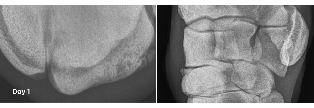
BACKGROUND and DIAGNOSTICS:
A 4year old flat-racing Thoroughbred mare presented 3/5 lame in the left forelimb with moderate effusion of the middle carpal joint and reaction to flexion. Diagnostic investigation revealed the source of pain to be emanating from the middle carpal joint and radiographs of both knees were taken.
RADIOGRAPHS:
These revealed that the horse had sustained a 3x3mm non-displaced slab fracture of the dorsoproximal aspect of the radial facet region of the third carpal bone. There was also evidence of sub-chondral bone disease in both third carpal bones.
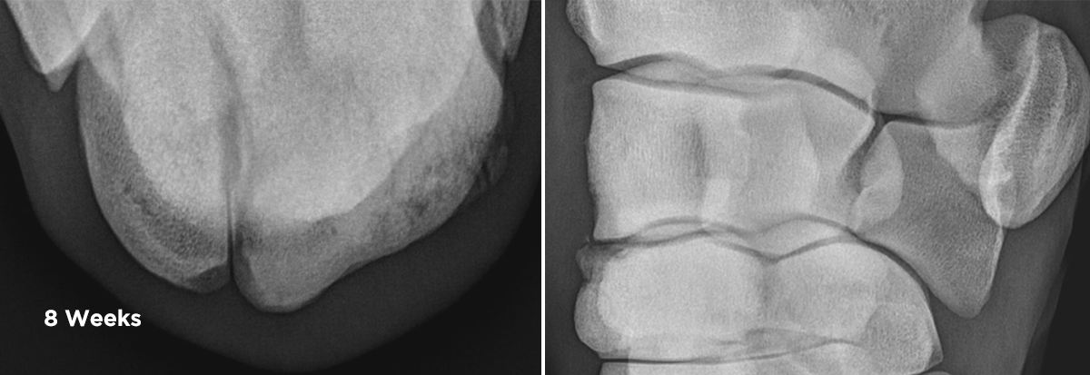
MANAGEMENT:
Due to financial constraints, the mare was managed conservatively with five days of oral phenylbutazone (an oral anti-inflammatory) and strict yard rest for eight weeks.
Follow up radiographs at this time revealed development of osteoarthritic changes within the left middle carpal joint. 2ml of Arthramid Vet was injected into the left middle carpal joint and a controlled rehabilitation program was instigated.
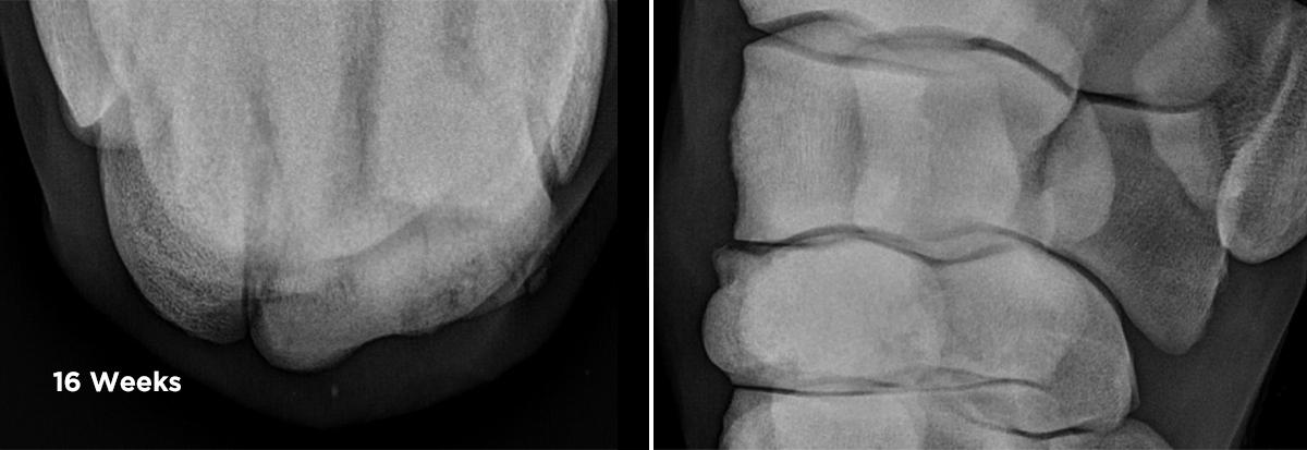
FOLLOW UP:
Further radiographs were taken eight weeks later and found there to be marked improvement in the osteoarthritic and third carpal bone changes and adequate healing of the fracture. Clinically, the horse was not lame, nor was there any reaction to flexion of the left knee nor effusion of the middle carpal joint.
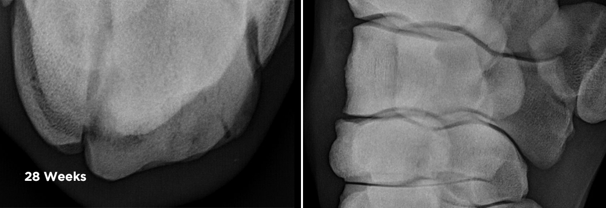
The horse re-entered a full training program with further radiographs taken three months later showing stabilisation of the left middle carpal joint and the horse displaying no signs of lameness or joint dysfunction.
Note: there are four pairs of rads which correlate to week 1, week 8, week 16 and week 28. The time signature on them is done is chronological order;